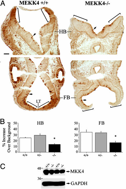
Fig. 4.
Reduced active MKK4 during neural tube closure in MEKK4-/- mice. (A) Immunostaining for phospho-MKK4 at E9.5 in the HB (Upper) and FB (Lower). Wild-type mice (Left) had strong staining in the neuroepithelial layers of dorsal HB and FB surrounding the LT. MEKK4-/- mice (Right) showed reduced staining in these areas. Bracketed regions were areas analyzed for quantification in Fig. 3B. Enriched staining in cells lining the HB and FB ventricles (arrows) was observed in both genotypes. (B) Quantification (described in Methods) of phospho-MKK4 staining in the HB and FB. MEKK4-/- mice showed significant decreases in the intensity of staining in both the HB and FB neuroepithelium compared with MEKK4+/+ or MEKK4+/- mice. *, P < 0.05 (ANOVA). (C) Western blot for total MKK4 at E9.5 showed comparable levels between MEKK4+/+ and MEKK4-/- mice. GAPDH was used as a loading control. (Bar, 50 μm in A.)

