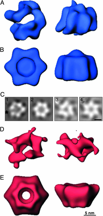
Fig. 2.
Three-dimensional structures of gp41 and gp61 complexes derived from electron microscopic images negatively stained with methylamine vanadate. Views are shown down the axis perpendicular to the plane of each ring (Left) and of a side view resulting from a 90° rotation of the en face view about its horizontal axis (Right). Each reconstruction was filtered to its resolution limit of 27 Å and is shown at a threshold corresponding to 100% of the mass of six copies of each protein and one of the 45-mer ssDNA. (A) The gp41 assembly with no rotational symmetry applied. (B) The gp41 assembly calculated with imposition of sixfold rotational symmetry. (C1–C4) En face projections corresponding to reconstruction of gp41 with no applied symmetry, with an asterisk marking the observed open interface of the hexamer (1), gp41 with sixfold rotational symmetry (2), gp61 with threefold rotational symmetry (3), and gp61 with sixfold rotational symmetry (4). (D) The gp61 assembly with no rotational symmetry applied. (E) The gp61 assembly calculated with imposition of sixfold rotational symmetry.

