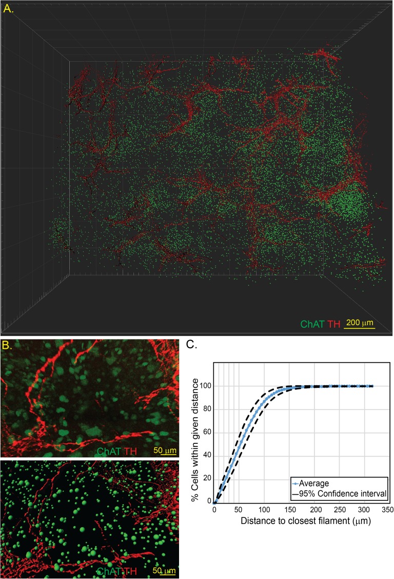
Fig 2. Three-dimensional reconstruction of splenic neuro-immune interactions by CLARITY.
Spleens from ChAT-GFP mice were subjected to CLARITY and imaged by two-photon microscopy (A) allowing for individual cells and neural surfaces to be identified and modelled (B). Distances of ChAT+ lymphocytes to the nearest TH+ axons was determined and quantified (C). Representative images from n = 6 animals.

