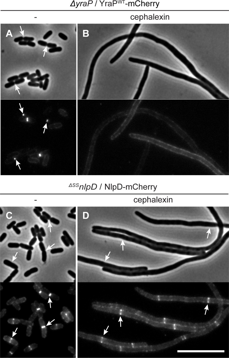
Fig 9. Localization of YraP or NlpD in cephalexin-treated cells.
Overnight cultures of (A-B) MT140 (ΔyraP) harboring the integrated construct attλMT197 (Plac::yraP-mCherry) or (C-D) MT47 (ΔSSnlpD) harboring the integrated expression construct attHKNP20 (Plac::nlpD-mCherry) were diluted in minimal M9-maltose medium supplemented with either 25μM (A-B) or 100μM (C-D) IPTG and grown at 30°C until mid-log. Cultures were then backdiluted into M9-maltose medium with the indicated IPTG concentration with or without 10μg/ml cephalexin as indicated. Cells were grown at 30°C to an OD600 of 0.2 before they were visualized on 2% agarose pads by phase contrast and fluorescence microscopy. Arrows indicate localization of the protein fusion to division sites. Bar = 10μm.

