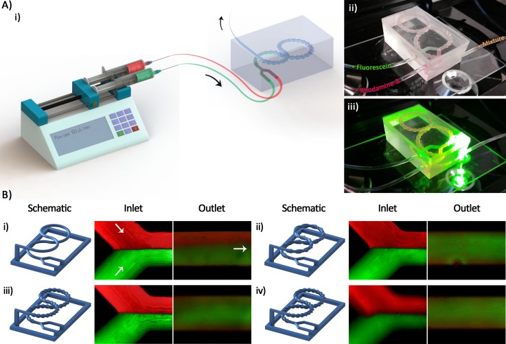
FIG. 8.
(a-i) A schematic illustration of the experimental setup: Two syringes of the same size containing the Rhodamine-B and Fluorescein solutions are connected to the fabricated micromixer through two sets of tubing of the same length. The solutions are pumped into the mixer using a syringe pump at the flow rate of 100 μl/min (Re = 1.58). ((a-ii) and (a-iii)) An optical picture of a micromixer placed on the microscope stage under illumination showing the condition of data acquisition. (b) Schematic of 3D printed wax structures as well as the microscopic images of the inlet and outlet of (b-i) mixer I, (b-ii) mixer II, (b-iii) mixer III, and (b-iv) mixer IV. Rhodamine-B (red) and Fluorescein (green) solutions at the same concentration were used for all of the mixers.

