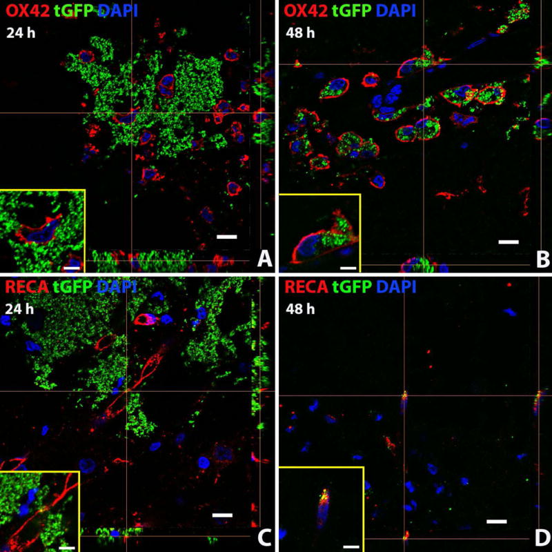
Figure 11.

Exogenous tGFP mitochondria are incorporated into different cell types in situ. tGFP mitochondria were injected into the naïve spinal cord. Representative z-stack images were taken from spinal cords at the 24 (A, C) or 48 hour (B,D) time points after injection. OX42 = microglia/macrophages, RECA = endothelial cells. Scale bars = 10μM. Images were taken with Nikon Ti confocal microscope.
