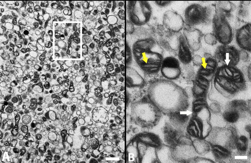
Figure 2.

TEM shows that the tGFP-labeled mitochondria have dense cristae (yellow arrows) and intact membranes (white arrows), indicative of healthy mitochondria. B is a higher magnification of boxed insert shown in A. Scale bars = 1 um (A), 200 nm (B).
