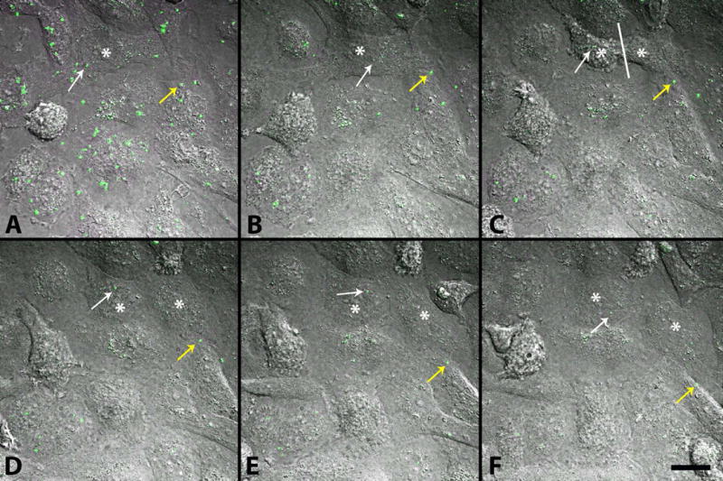
Figure 9.

Movement of exogenous tGFP mitochondria within PC-12 cells. After 10 μg tGFP mitochondria was added to naïve PC-12 cells and incubated at 37°C on a gently rolling platform for 2 hours, they were washed off and replaced with complete media before time lapse images were taken over a 12-hour period. Images represent beginning of imaging (A) separated by 100 minutes for each time frame (B–F). Mitochondria can be visualized moving within cells (yellow arrow). As a cell divides (white asterisk on mother cell in A and B, then on each daughter cell in C–F, with division occurring at white line in C), transplanted mitochondria can be seen throughout the division process, and are retained in the daughter cells (white arrow). Images were taken using Nikon Ti confocal microscope. Scale bar = 20μM.
