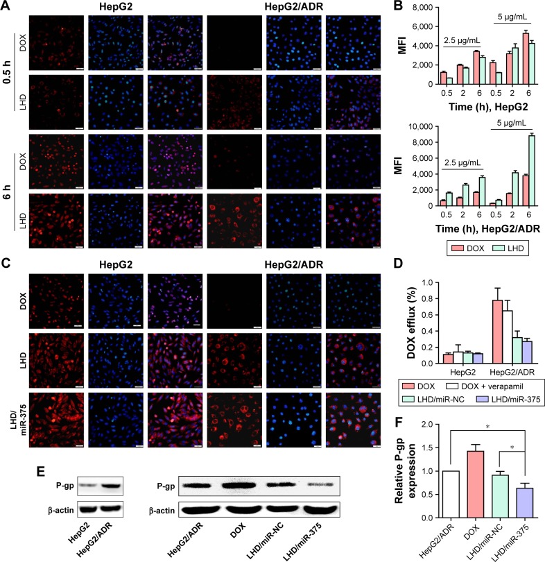
Figure 3.
Uptake and retention of LHD in HepG2 and HepG2/ADR cells.
Notes: (A) Uptake of LHD in HepG2 and HepG2/ADR cells determined by fluorescence microscopy. Cells were incubated with DOX (5 μg/mL) or LHD (5 μg/mL). (B) Uptake of LHD in HepG2 and HepG2/ADR cells determined by flow cytometry. Cells were incubated with DOX (2.5 or 5 μg/mL) or LHD (2.5 or 5 μg/mL). (C) Retention of LHD/miR-375 in HepG2 and HepG2/ADR cells determined by fluorescence microscopy. HepG2 and HepG2/ADR cells were incubated with DOX (5 μg/mL), LHD (5 μg/mL DOX), or LHD/miR-375 (5 μg/mL DOX and 100 nM miR-375) for 4 h and then cultured with medium without drugs for another 24 h. (D) DOX efflux determined by flow cytometry. HepG2 and HepG2/ADR cells were treated with DOX (5 μg/mL), DOX + verapamil (DOX 5 μg/mL and verapamil 10 μM), LHD/miR-NC (DOX 5 μg/mL and 100 nM miR-NC), or LHD/miR-375 (5 μg/mL DOX and 100 nM miR-375). The nuclei were counterstained by 4,6-diamidino-2-phenylindole. (E) Western blotting assay for P-gp protein expression after DOX, LHD/miR-NC, or LHD/miR-375 treatment in HepG2/ADR cells. (F) P-gp protein level by quantifying the Western blotting bands. P-gp level in untreated cells was standardized to 1. Data are expressed as mean ± standard error of the mean of three independent experiments. *P<0.05.
Abbreviations: DOX, doxorubicin hydrochloride; LHD, lipid-coated hollow mesoporous silica nanoparticles containing doxorubicin hydrochloride; P-gp, P-glycoprotein; MFI, mean fluorescence intensity; NC, negative control.

