. 2017 Jul 28;6:e25885. doi: 10.7554/eLife.25885

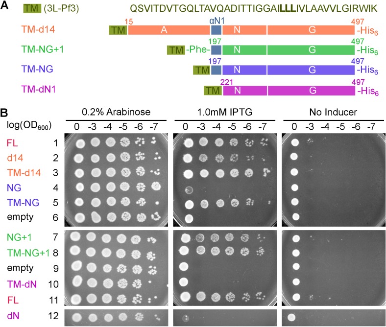
Figure 8. Pre-organization into the Stable mode disrupts FtsY function in vivo.

(A) The sequence of the 3L-Pf3 TMD anchor and domain structures of membrane-tethered FtsY mutants (TM-FtsY) used in the in vivo assay. (B) Frogging assays were carried out to test the ability of mutant FtsYs to complement the loss of genomic FtsY, as described in Methods. Replicates of the data are shown in Figure supplement.

DOI: http://dx.doi.org/10.7554/eLife.25885.021

Figure 8.

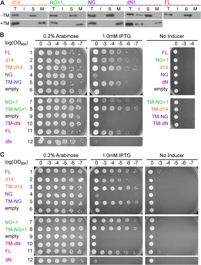
Figure 8—figure supplement 1. Cell fractionation analyses and replicates of the cell growth assay.

Figure 8—figure supplement 1.

(A) Cell fractionation analysis to test the effectiveness of membrane tethering by the 3L-Pf3 motif. FtsY truncation mutants without (-TM) or with (+TM) the 3L-Pf3 fusion were expressed and fractionated as described in Methods. Proteins in total (T), inclusion body (I), soluble (S), and membrane (M) fractions were detected via Western-blotting against their C-terminal His6-tags. (B, C) Two independent replicates of the frogging assays (Figure 8B) to test the ability of mutant FtsYs to complement FtsY depletion.