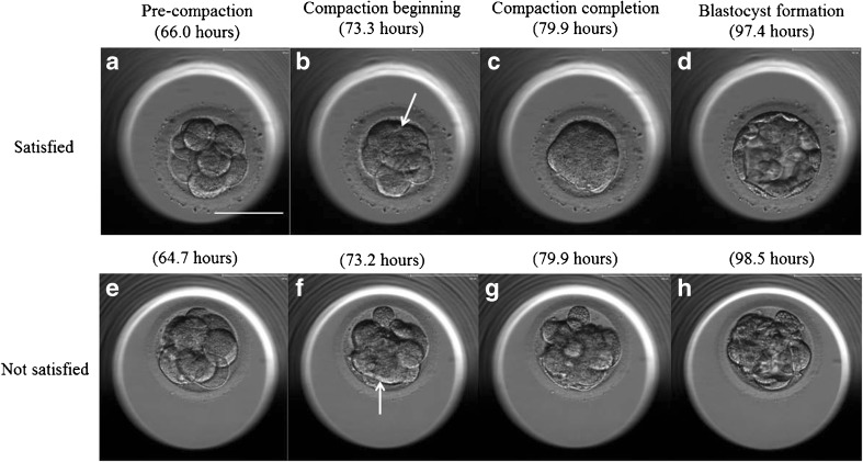
Fig. 1.
In vitro development of a–d “satisfied” and e–h “not satisfied” embryos. a, e Pre-compaction, b, f compaction beginning, c compaction completion, d blastocyst, g incomplete compaction, and h incomplete blastocyst. Culture times are shown in each picture. The arrows show the obscured intercellular boundaries by the merging of blastomeres. Scale bar 100 μm

