
Figure 5.
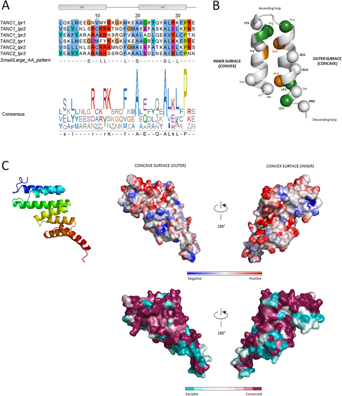
TPR repeat overview and TANC2 TPR model. (A) Consensus sequence repeat pattern of the TANC TPR domain and related sequence logo. Secondary structure is shown above the alignment: two alpha helices (grey shapes) connected by a loop (black line). Below the alignment, pattern of conserved small/large residues typical of TPR modules is reported: S indicates small residues, L for large residues. Residues that match the consensus are reported in upper case. (B) Graphic representation of repeats structure in TANC proteins. Conserved positions of TPR consensus pattern are reported in the diagram (spheres). Residues that match the consensus are reported in bold. Conserved small-large residue pattern is also represented: dark green for large residues and orange for small residues. (C) Cartoon of TANC2 TPR domain model is coloured from N-terminus (blue) to C-terminus (red). Electrostatic properties of concave and convex surfaces are shown: negative charges in blue and red charges in red. ConSurf analysis of turn-loop surfaces and connecting-loop surfaces, colour code from unconserved (cyan) to conserved (purple) residues.