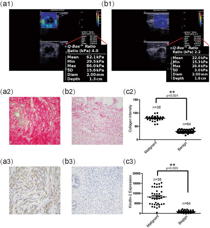
Figure 2.

Evaluation of malignant and benign breast nodules by SWE and by histopathological staining for collagen and Kindlin-2 expression. (a1) (b1) Representative SWE images of malignant and benign breast nodules. (a2) (b2) Representative images from Sirius red staining (×200) show expression of collagen in malignant and benign breast nodules, respectively. (c2) Quantitative analysis shows that the average intensity of collagen in malignant breast nodules was significantly higher than that in benign breast nodules. (a3) (b3) Representative images of immunohistochemical staining (×200) show expression of Kindlin-2 in malignant and benign breast nodules, respectively. (c3) Quantitative analysis shows that the average expression (by integrated optical density of positive reactions) of Kindlin-2 in malignant breast nodules was significantly higher than that in benign breast nodules. Values are means ± SD. Comparisons between two groups were made using Student’s t-test. *p < 0.05, **p < 0.01. SWE, shear wave elastography; SD, standard deviation.
