Figure 5.

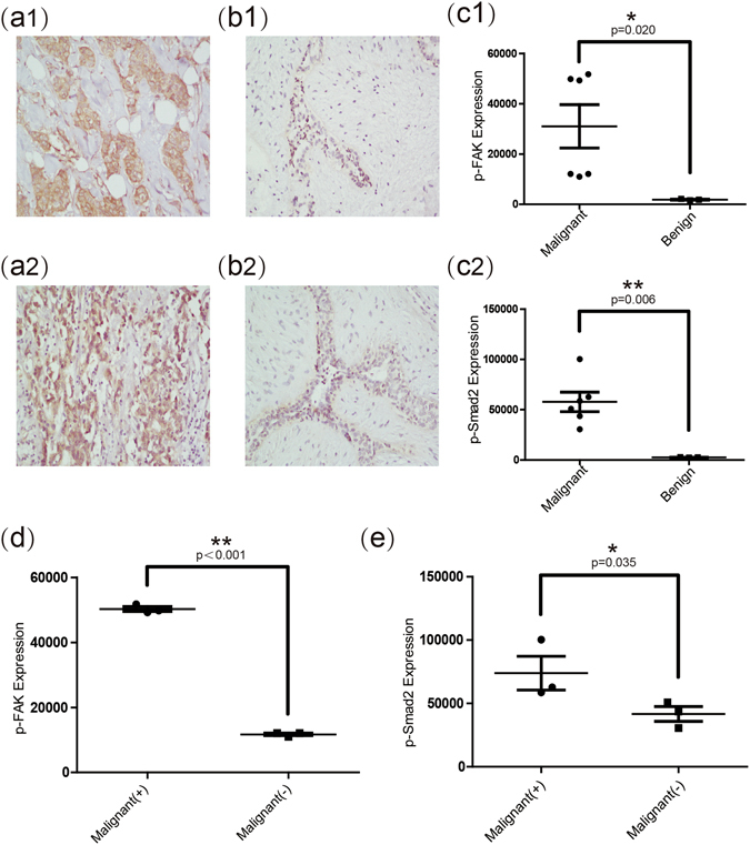
Expression levels of p-FAK and p-Smad2 in malignant nodules with or without lymph nodes metastasis, and in benign nodules. (a1) (b1) Representative images of immunohistochemical staining (×200) show expression of p-FAK in malignant and benign breast nodules. (c1) Quantitative analysis shows that the average expression (by integrated optical density of positive reactions) of p-FAK in malignant breast nodules was significantly higher than that in benign breast nodules. (a2) (b2) Representative images of immunohistochemical staining (×200) show expression of p-Smad2 in malignant and benign breast nodules. (c2) Quantitative analysis shows that the average expression (by integrated optical density of positive reactions) of p-Smad2 in malignant breast nodules was significantly higher than that in benign breast nodules. (d) The p-FAK expression of malignant breast nodules with lymph node metastasis was significantly higher than that without lymph node metastasis. (e) The p-Smad2 expression of malignant breast nodules with lymph node metastasis was significantly higher than that without lymph node metastasis. Values are means ± SD. Comparisons between two groups were made using Student’s t-test. *p < 0.05, **p < 0.01. SD, Standard deviation.
