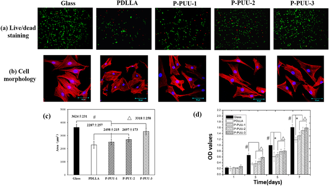
Figure 5.

Morphology and proliferation of osteoblasts on different substrates: (a) Live/dead staining for cell after seeding of 24 h, green fluorescence indicating cells alive while the red visualizing dead cells; (b) Morphology and of osteoblasts on glass, PDLLA, P-PUU-1, P-PUU-2, and P-PUU-3 by CLSM at 24 h; (c) cell spreading areas (n = 200) after seeding of 24 h; (d) Cell proliferation by MTT at 4, 7, 14 and 21 days (# means P < 0.05, compared with glass groups; * means P < 0.05, compared with PDLLA groups; Δ means P < 0.05, compared with P-PUUs groups).
