Figure 3.

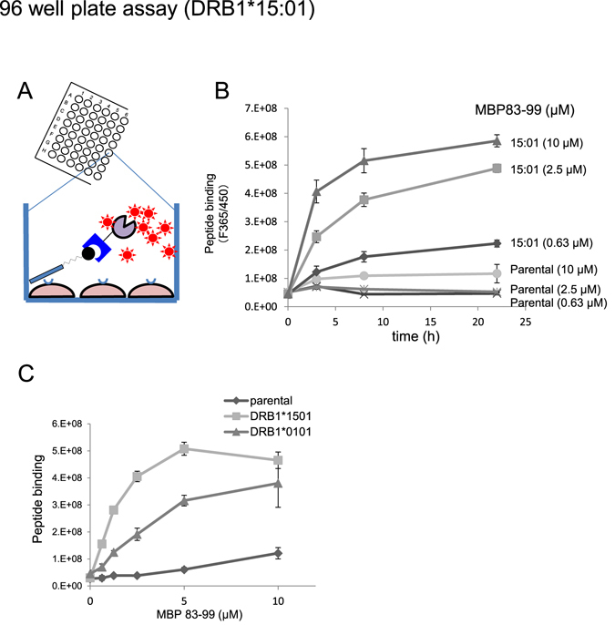
Detection and screening system of antigen peptide binding to HLA on 3T3 cells in 96 well plate. (A) Scheme of the detection of HLA-bound peptides on cells in 96 well plates. HLA expressing cells are incubated with biotinylated antigen peptides, and bound peptides are probed with SA-β-gal, followed by enzyme reaction with chromogenic (ONPG) or fluorogenic substrate (4MUG). (B) Detection of HLA-bound peptides on 3T3 cells using fluorogenic substrate (4MUG) in 96 well plates. Two 96-well plates of DR15-expressing cells were incubated with MBP83-99 for 6 h, fixed with glutaraldehyde, and subjected to blocking with a blocking solution as detailed in the methods section. Thereafter, biotinylated peptides in one plate were probed with low concentration of SA-β-gal (3,000-fold dilution) and detected by using 4MUG. The other plate was analysed using a chromogenic substrate (ONPG) in Supplementary Fig. 3. (C) Time and concentration dependence of MBP83-99 binding to DR15-expressing 3T3 cells. The assay was conducted as in (B). In either figure, values shown are mean ± intra-assay deviation expressed as SD from 3 wells in one set of representative experiments.
