
Figure 1.
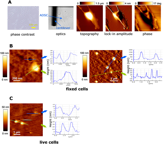
Morphology of protrusions and pits on ADSC membranes by AFM. (A) Characterization of cultured ADSCs under optic phase contrast, under direct light at the AFM set-up and under different AFM analyses (topography, lock-in amplitude and phase). (B) AFM measurements of fixed cells in two 10 × 10 µm fields. Medium sized (height: 100–200 nm) and small sized (height 20–100 nm) protrusions bulge on the surface. Also, circular, crater-like depressions with raised rims are present (measurements of the actual depth of pits is limited by probe tip constraints). (C) In living cell preparations at 37 °C, similar buddings and depressions were observed. We hypothesize that bulges are the origin of shedding vesicles and depressions are the surface appearance of membrane-fused MVBs for exosomes release.