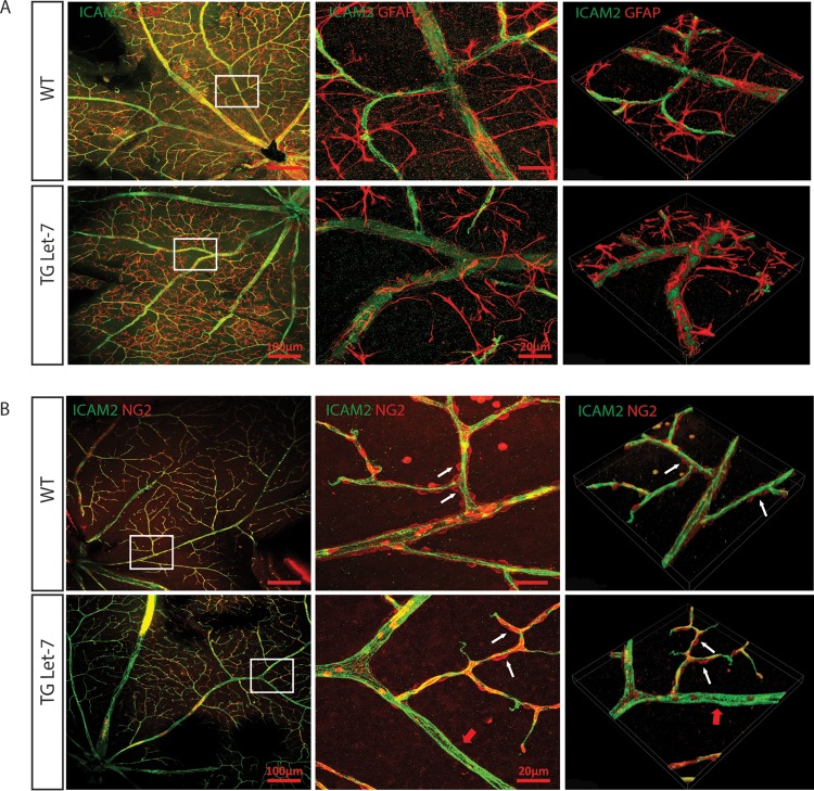
FIG 6.
let-7 transgenic mice have normal astrocyte coverage but defective pericyte coverage. (A) Normal astrocyte coverage in 4-month-old let-7-Tg mice compared to WT mice, as shown by ICAM-2 and GFAP costaining. The images in the middle panels represent the boxed areas in the left panels. The images in the right panels are three-dimensional constructions of the representative confocal images. (B) Defective pericyte coverage in 4-month-old let-7-Tg mice compared to WT mice, as shown by ICAM-2 and NG2 costaining. The images in the middle panels represent the boxed areas in the left panels. The images in the right panels are three-dimensional constructions of the representative confocal images. White arrows indicate the pericyte coverage in the arterioles, while red arrows indicate the lack of pericyte coverage in some regions of the let-7-Tg mice.

