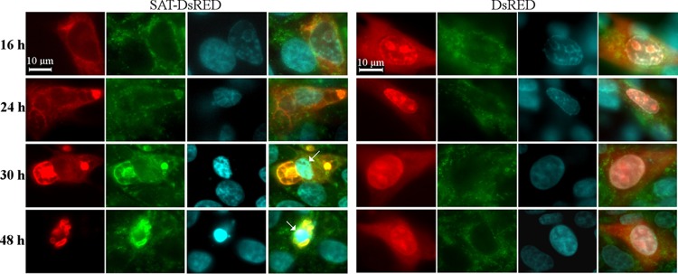
FIG 9.
Morphological changes of the ER in SAT-DsRed fusion protein-expressing PT cells. Cells were transfected with a SAT-DsRed-expressing plasmid and, as a control, with a DsRed (red)-expressing plasmid. The calreticulin was labeled by anticalreticulin monoclonal antibody (green), and the cell nuclei (blue) were visualized by Hoechst staining. The cells were fixed at different times after transfection. Arrows, apoptotic nuclei.

