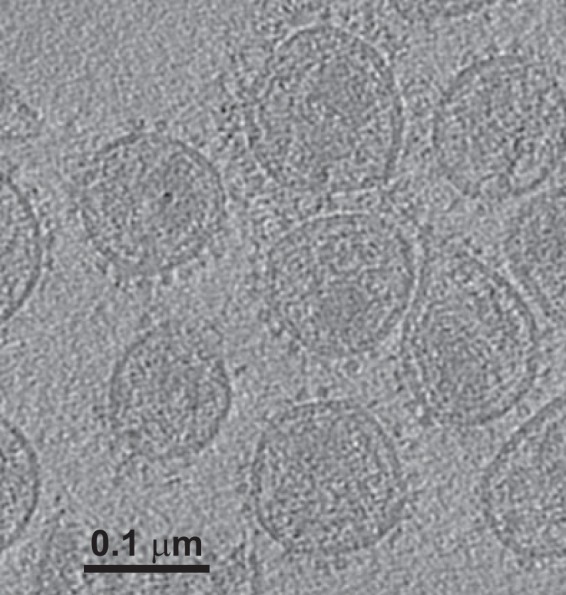
FIG 1.

Tomogram of SIV. The image shows a single section from one tomogram of strain SIVmac239/251 tail/Supt-CCR5 CL.30, revealing envelope spikes on the virion surface. These virions were preincubated with sCD4 as well as MAb 36D5. Ligands are difficult to see unless they lie within the section plane.
