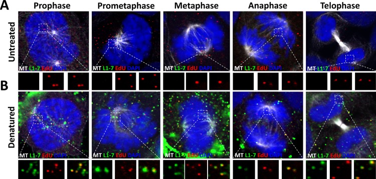
FIG 4.
The linear epitope of the 33L1-7 antibody is hidden in the L1 protein associated with condensed chromosomes. (A and B) At 24 hpi, HaCaT cells infected with EdU-labeled pseudovirus were fixed, permeabilized, and treated with or without with Click-iT reaction buffer without dye. Next, the cells were incubated with mouse MAb 33L1-7 antibody (green) and AF488-conjugated anti-α-tubulin (white). Lastly, the cells were treated with Click-iT reaction buffer with AF555 dye (red) to stain the EdU-labeled pseudogenome and mounted in DAPI (blue). Note that colocalization of the EdU puncta and L1 protein is denoted as a yellow color.

