FIG 8.

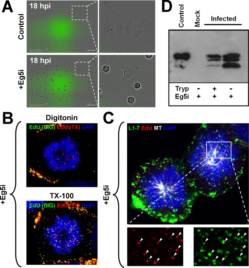
Some full-length L1 protein accompanies the viral genome to the nucleus. (A) HeLa cells were infected with HPV16 pseudovirus with or without 1.5 μM Eg5 inhibitor III (Eg5i) and tracked via live-cell imaging using the IncuCyte Zoom for 48 h. Note that the images are depicted at 18 hpi. (B) HaCaT cells were infected with EdU-labeled HPV16 pseudovirus in the presence of 1.5 μM Eg5i. The cells were fixed at 24 hpi and permeabilized with either digitonin at 0.625 μg/ml or 0.5% TX-100 and then treated with AF555 (green) in Click-iT reaction buffer. The cells were permeabilized again with 0.5% TX-100 and treated with AF647 (red) in Click-iT reaction buffer. Lastly, the cells were mounted in DAPI (blue). (C) HaCaT cells were infected with EdU-labeled pseudovirus and treated with 1.5 μM Eg5i. At 24 hpi, the cells were fixed and permeabilized in 0.5% TX-100. Next, the cells were treated with AF555 (red) in Click-iT reaction buffer, followed by incubation with AF488-conjugated anti-α-tubulin (white) and MAb 33L1-7 (green). Lastly, the cells were mounted in DAPI (blue). Note the colocalization between EdU and L1 signal denoted by white arrows. (D) HeLa cells were infected with HPV16 pseudovirus in the presence of 1.5 μM Eg5i. Cells were trypsinized for 2 min, and monoastral cells were collected. Next, the cells were treated with 15 μl of 0.25% trypsin for 1 h at 37°C. The cells were lysed by passage through a 1-ml syringe with a 25-gauge needle 40 times. Cell lysates were incubated for 1 h at 37°C once more, the trypsin was inactivated, and the samples were analyzed by Western blot analysis with a cocktail of HPV16 L1-specific mouse MAbs (IID5, 33L1-7, and 312F).
