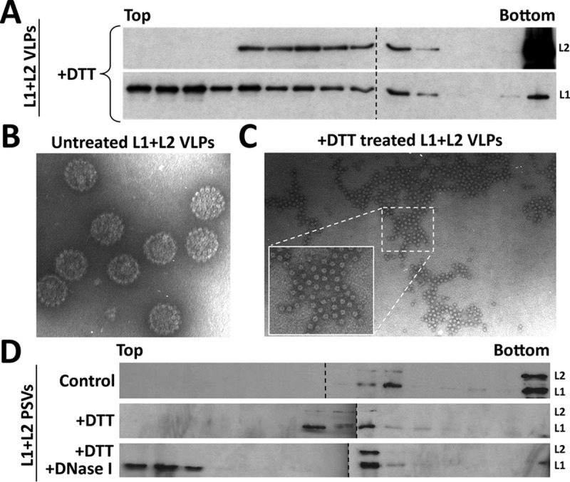
FIG 9.

L1 protein interacts with the viral DNA within the capsid. (A) Purified DNA-free HPV16 L1/L2 VLPs were incubated with 20 mM DTT for 15 min at room temperature. Samples were layered on the top of a 20 to 60% sucrose linear gradient and subjected to ultracentrifugation. Collected fractions were analyzed by Western blotting for specific detection of the L1 and L2 proteins using MAbs. (B and C) Cesium chloride-purified DNA-free HPV16 L1/L2 VLPs were analyzed by electron microscopy with or without 20 mM DTT treatment. (D) HPV16 L1/L2 pseudoviruses were incubated with 10 mM MgCl2, with or without 20 mM DTT, and with or without 2 U of DNase I for 30 min at 37°C. Samples were layered on the top of a 20 to 60% sucrose linear gradient and subjected to ultracentrifugation. Collected fractions were analyzed by Western blotting for the specific detection of L1 and L2 proteins using MAbs.
