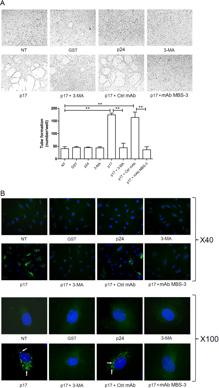
FIG 2.
Inhibition of autophagy with 3-MA decreases p17-mediated lymphangiogenesis. (A) LN-LECs were starved for 16 h and then left unstimulated or stimulated or with 10 ng/ml of GST, p24, or p17 in complete medium. LN-LECs were starved for 16 h in the presence or absence of 3-MA (5 mM), as indicated. In selected experiments, LN-LECs were stimulated with p17 (10 ng/ml) after preincubation of the viral protein with 1 μg/ml of MAb to p17 (MAb MBS-3) or an unrelated control MAb (Ctrl MAb) for 30 min at 37°C. Pictures were taken after 12 h of culture on growth factor-reduced Cultrex BME (magnification, ×10) and are representative of three independent experiments with similar results. Values reported for tube formation are the means ± SDs of three independent experiments with similar results. Statistical analysis was performed by one-way ANOVA; a Bonferroni posttest was used to compare data (**, P < 0.01). (B) LN-LECs were nucleofected with EGFP-LC3 plasmid, and 24 h after nucleofection cells were starved for 16 h and then left unstimulated or stimulated with 10 ng/ml of GST, p24, or p17 for 3 h in complete medium. When indicated, LN-LECs were starved for 16 h in the presence or absence of 3-MA (5 mM). In selected experiments, LN-LECs were stimulated with p17 (10 ng/ml) after preincubation of the viral protein with 1 μg/ml of MAb to p17 (MAb MBS-3) or an unrelated control MAb (Ctrl MAb) for 30 min at 37°C. The images display LC3 signals in green and cell nuclei in blue. Magnification is as indicated on the figure. Green-positive punctate structures were counted in order to quantify relative levels of autophagy. The percentage of positive cells was calculated for 10 independent fields at a magnification of ×40. A total number of 29 ± 10 cells per microscope field was counted, and the percentage of LC3-positive punctate cells under p17 stimulation was found to be 38% ± 3%. White arrows indicate dot-like LC3+ structure formation. NT, not treated.

