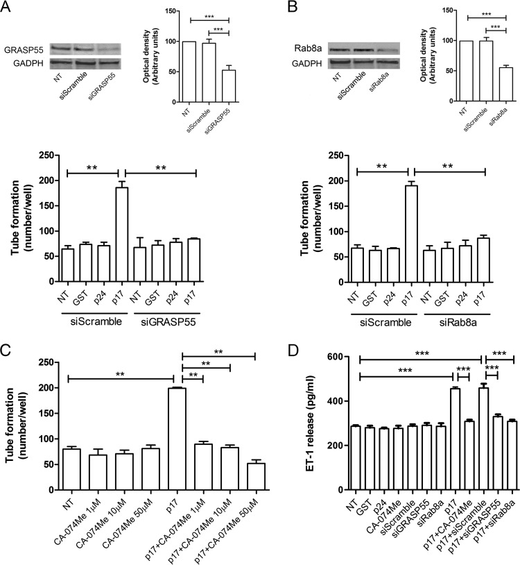
FIG 7.
Mechanistic insight into the prolymphangiogenic activity of p17. LN-LECs were nucleofected with siGRASP55, siRab8a, and siScramble. (A and B) Immunoblotting analysis was performed on LN-LECs at 24 h after GRASP55 (A, upper panel) or Rab8a (B, upper panel) siRNA nucleofection. Blots from one representative experiment of three with similar results are shown. Values reported for GRASP55 or Rab8a knockdown are the means ± SDs of three independent experiments with similar results. Twenty-four hours after nucleofection, cells were serum starved for 16 h, and then a tube formation assay was performed with either untreated LN-LECs or with siScramble-nucleofected LN-LECs or LN-LECs silenced for GRASP55 (A, lower panel) or Rab8a (B, lower panel) with 10 ng/ml of GST, p24, or p17 in complete medium. (C) Tube formation assay of LN-LECs serum starved for 16 h in the presence or absence of 1, 10, and 50 μM CA-074Me (cathepsin-β inhibitor) and then seeded on growth factor-reduced Cultrex BME-coated wells and either untreated or treated with p17 (10 ng/ml) in complete medium. Values reported for tube formation are the means ± SDs of three independent experiments with similar results. (D) LN-LECs were serum starved in the presence or absence of 50 μM CA-074Me. In some experiments, LN-LECs were nucleofected with siScramble, siGRASP55, or siRab8a, and 24 h after nucleofection, cells were serum starved for 16 h. After starvation, LN-LECs were cultured in complete medium either unsupplemented or containing 10 ng/ml of GST, p24, or p17. After 6 h of culture, supernatants were collected and analyzed by a standard quantitative ELISA. Bars represent the means ± SDs of triplicate samples. Statistical analysis was performed by one-way ANOVA; a Bonferroni posttest was used to compare data (**, P < 0.01; ***, P < 0.001). NT, not treated.

