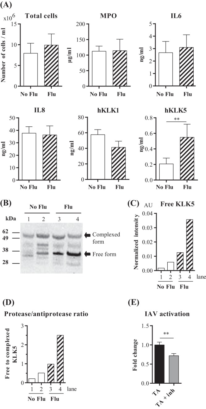
FIG 5.

KLKs in lower respiratory tract secretions. (A) Total cell numbers and concentrations of myeloperoxidase, inflammatory mediators (IL-6 and IL-8), and KLKs were measured in tracheal aspirate samples from control patients (No Flu) (n = 18) and influenza patients (Flu) (n = 17) in an intensive care unit. Means ± standard errors of the means are shown. **, P < 0.01, as determined by a Mann-Whitney U test. (B) Western blot analysis of KLK5 in tracheal aspirates from patients in an intensive care unit. Lanes from the gel images were cut and spliced together with Adobe Photoshop CS 5.1 to aid in band comparison. Numbers at the left refer to molecular masses, in kilodaltons. (C) Densitometric analysis of the Western blot data shown in panel B. The values (AU, arbitrary units) represent the signal intensity normalized against the total protein amount in each lane assessed by Ponceau S staining after membrane transfer. (D) Ratio of free to bound KLK5. (E) MDCK cells were infected with influenza A/Scotland/20/74 virus particles that had been pretreated with supernatants (n = 9) of human tracheal aspirates (TA) in the absence or in the presence of a selective KLK5 inhibitor (TA + Inh) for 16 h at 37°C. Proportions of infected MDCK cells were determined by flow cytometry using an anti-NP-FITC antibody. Results are expressed as fold changes of values obtained without an inhibitor. Data were analyzed by a Mann-Whitney U test (**, P < 0.01).
