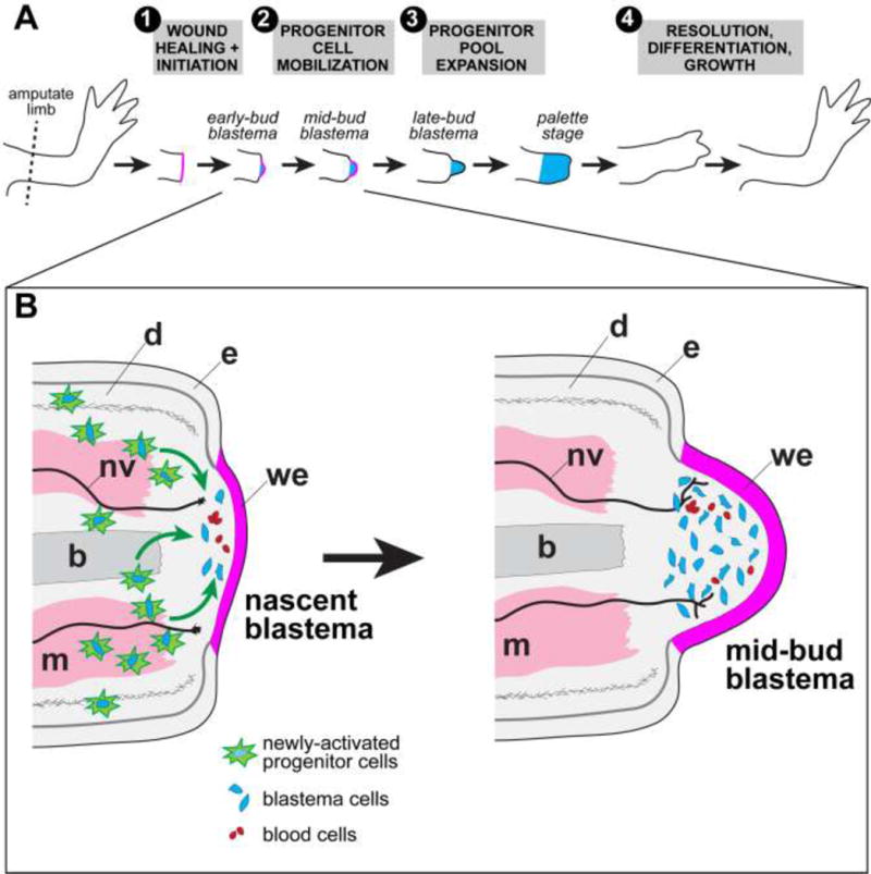
Figure 2. Outline of cellular events during limb regeneration.

(A) General progression from unamputated to fully regenerated. (1) Immediately following amputation (within ~24 hours), a thin wound epidermis (magenta) forms across the cut stump via migration of stump epidermal cells. Wound epidermis thickens as cells within it proliferate. (2) A visible bump, called a blastema (blue), forms beneath the wound epidermis. Blastema cells are derived from activated progenitor cells within various stump tissues that migrate to the tip. (3) Blastema cells proliferate to expand the progenitor pool. (4) The initial regeneration response resolves, cells begin to undergo differentiation, and the limb continues to grow to the appropriate size. (B) Early steps are shown in more detail in inset. Architectures of tissues such as bone and muscle are locally deconstructed near the amputation plane and are therefore shown as jagged. Newly-activated progenitor cells, which give rise to future blastema cells, are depicted with bright green starburst outlines. These cells are cued to re-enter the cell cycle and some fraction of them presumably migrate to the space immediately below the wound epidermis. Blood cells, both red and white, are intermingled with blastema cells. A “nascent blastema” is equivalent to very early-bud stage blastema in other literature. Noted are: epidermis (e), wound epidermis (we), dermis (d), bone (b, medium gray), muscle (m, pink), nerves (nv, black). Migration of newly-activated progenitor cells to the tip of the stump during blastema formation is implied by the green arrows.
