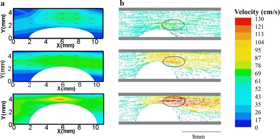
Fig. 5.

Velocity measured by Echo PIV and CFD in the systolic peak. a Velocity vectors measured by Echo PIV, red arrows represented the flow directors. An obvious vortex occurred in the back of 70% stenosis phantom. The peak blood velocity increased when increasing the stenosis degree. b Velocity vectors obtained by CFD. The dotted portion indicated the largest velocity areas
