Fig. 7.

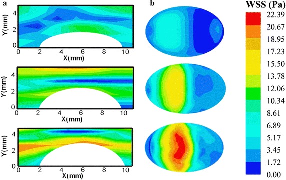
SS measured by Echo PIV and CFD in the systolic peak. a SS distribution in the central plane measured by Echo PIV in three stenosis phantoms. b WSS distribution in outer wall measured by CFD

SS measured by Echo PIV and CFD in the systolic peak. a SS distribution in the central plane measured by Echo PIV in three stenosis phantoms. b WSS distribution in outer wall measured by CFD