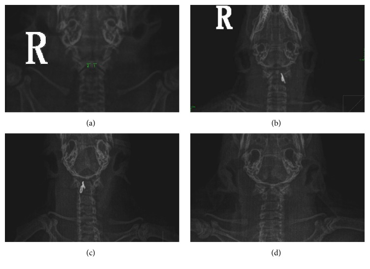
Figure 1.
X-ray imaging to judge atlantoaxial disorder rats. (a) The atlantoaxial joints of normal rats under X-ray system. (b) The implanted fixture was inserted into the left atlantoaxial joint. (c) The implanted fixture was inserted into the right atlantoaxial joint. (d) X-ray images after the implant was removed.

