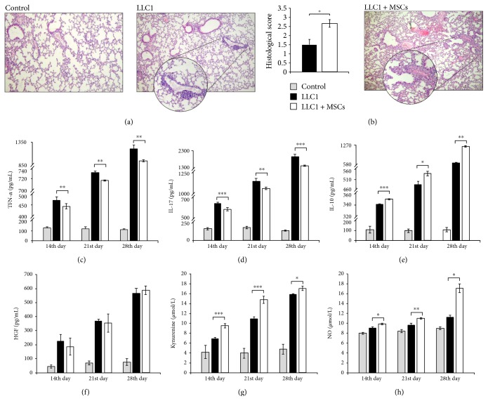
Figure 1.
MSCs promote lung cancer metastasis. (a) Representative H&E stained mouse lungs obtained at the 28th day of the experiment. H&E staining images of liver tissue samples are shown at the same magnifications (×100). (b) Histological score of lung tissue determined at the 28th day of the experiment. (c) Serum concentrations of TNF-α, (d) IL-17, (e) IL-10, and (f) HGF measured at the 14th, 21st, and 28th days of the experiment. (g) The level of kynurenine and (h) NO in mouse sera at the 14th, 21st, and 28th days of the experiment. Data presented as mean ± SEM; n = 10 mice per experimental groups. ∗p < 0.05, ∗∗p < 0.01, and ∗∗∗p < 0.001.

