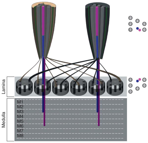
Figure 5.4.
Axonal targeting differences between outer and inner photoreceptors. Diagram representing two ommatidia sharing lamina cartridges. The axons from the six outer PRs from each ommatidium turn 180° and project to six different cartridges present in the lamina neuropil present directly underneath the retina. R1–R6 positions within the lamina represent a mirror image of the outer photoreceptor arrangement found in the retina. The R7 (magenta) and R8 (blue) axons bypass the lamina and project to layers M3 and M6 respectively in the adult medulla.

