
Figure 6.
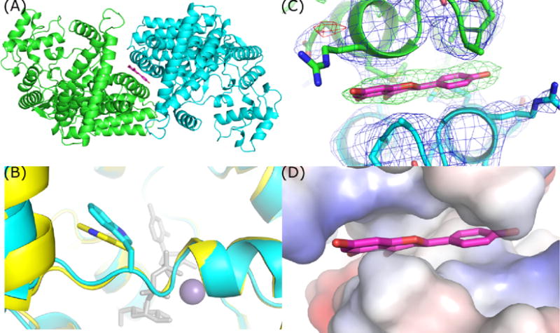
TcdB-GTD crystallized in an apo-like conformation contains two chains within the ASU. (A) Top-down view demonstrating the position of apigenin and non-crystallographic symmetry of the TcdB-GTD chains. (B) Overlay of apo TcdA-GTD (yellow) and TcdB-GTD (cyan) in an apo-like conformation. (C) 2mFo-DFc and Fo-Fc maps of apigenin site contoured at 1 and 4 sigma respectively. (D) Vacuum electrostatic model of the hydrophobic patch occupied by apigenin.