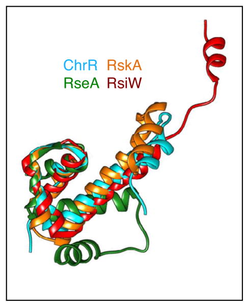
Figure 2.
Structural conservation of the ASD from ChrR, RseA, RskA, and RsiW. Structural alignments were performed using Swiss-PDBViewer 4.0 and displayed using Chimera [71]. The three-helix bundle is superimposable for all the anti-sigma factors, while the position of the fourth helix is variable. ChrR is shown in cyan, RseA in green, RskA in orange, and RsiW in red.

