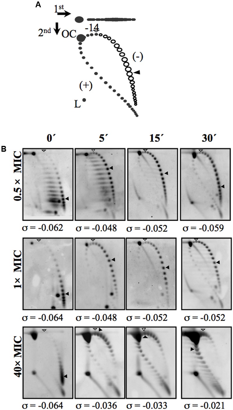
FIGURE 3.

Treatment with the GyrB inhibitor NOV causes relaxation and subsequent recovery of supercoiling levels. (A) Diagram showing plasmid pLS1 topoisomer distribution after two-dimensional electrophoresis in agarose gels run in the presence of 1 and 2 μg/ml chloroquine in the first and second dimensions, respectively. Arrows at the top left corner indicate the running direction of the first and second dimensions, respectively. OC, open circle; L, linear form. Negative supercoiled topoisomers are in white and positive supercoiled topoisomers in black. 2 μg/ml chloroquine introduces 14 positive supercoils. A white arrowhead indicates the topoisomer that migrated with ΔLk of 0 in the second dimension; it migrated with a ΔWr of –14 in the first dimension. A black arrowhead indicates the most abundant topoisomer. (B) pLS1 topoisomer distribution after different NOV treatments. Samples were taken before the addition of the drug (time 0 min) and at the times indicated. The corresponding supercoiling density (σ) value is indicated below each autoradiogram. Taken from Ferrándiz et al. (2010), with modifications.
