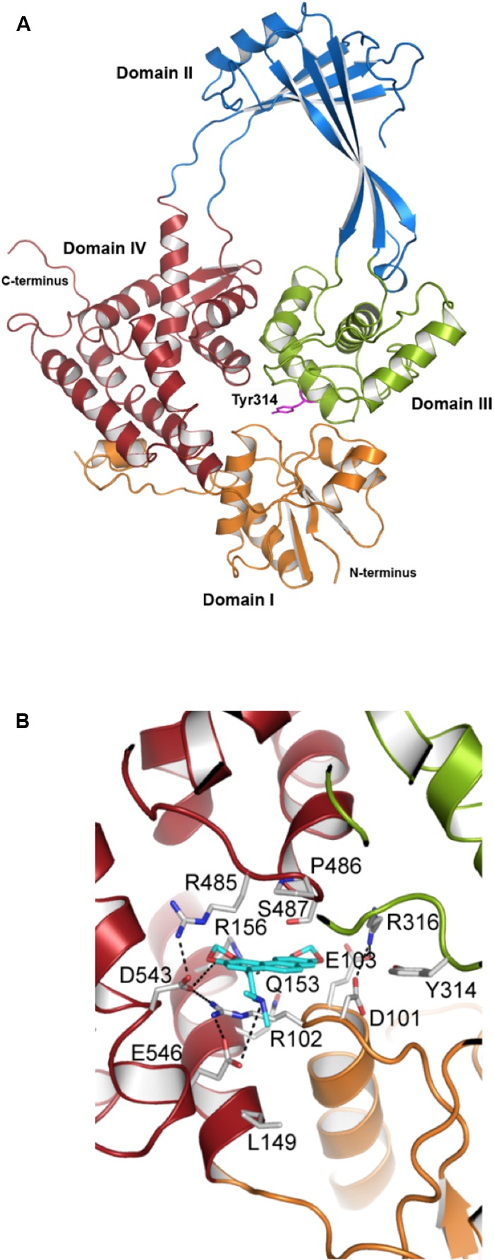
FIGURE 6.

Structural modeling of the interaction of N-methyl SCN with S. pneumoniae topoisomerase I. (A) Modeling of the 67 kDa fragment of Topo I, showing domains I–IV and the catalytic Tyr314. (B) N-methyl-SCN (in blue) bound to the nucleotide-binding site of Topo I. Hydrogen bonds and salt-bridge interactions are indicated by dashed lines. Taken from García et al. (2011), with modifications.
