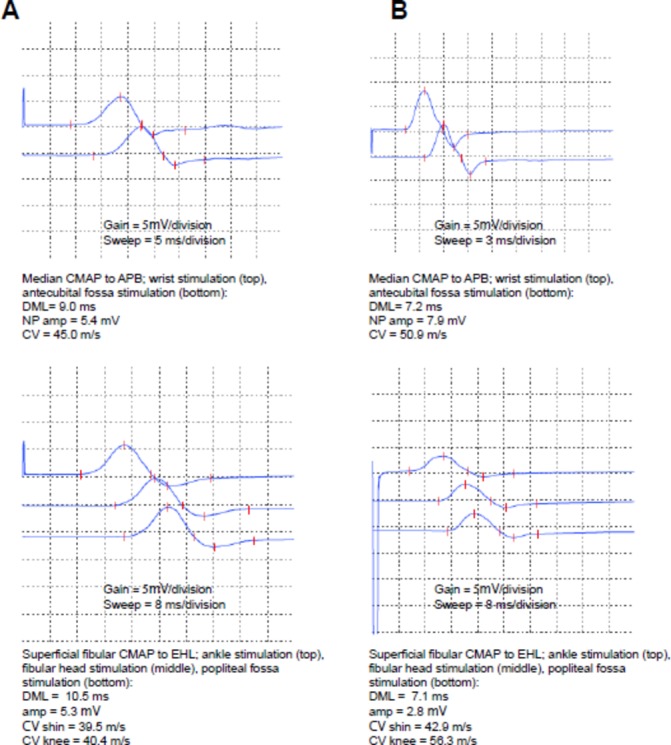
Figure 2.

Compound muscle action potentials for the median (top) and superficial fibular (bottom) nerves at the time of initial presentation (A) and at follow-up (B). Gain and sweep speed are highlighted below the individual waveforms for reference. Note the difference in sweep speed for the median study (top panels), to explain the difference in waveform morphology between A and B. Parameters of the compound muscle action potentials are listed below the respective waveforms for reference. APB, abductor pollicis brevis; CMAP, compound muscle action potential; CV, conduction velocity; DML, distal motor latency; EDB, extensor digitorum brevis; NP amp, negative peak amplitude.
