Figure 5.

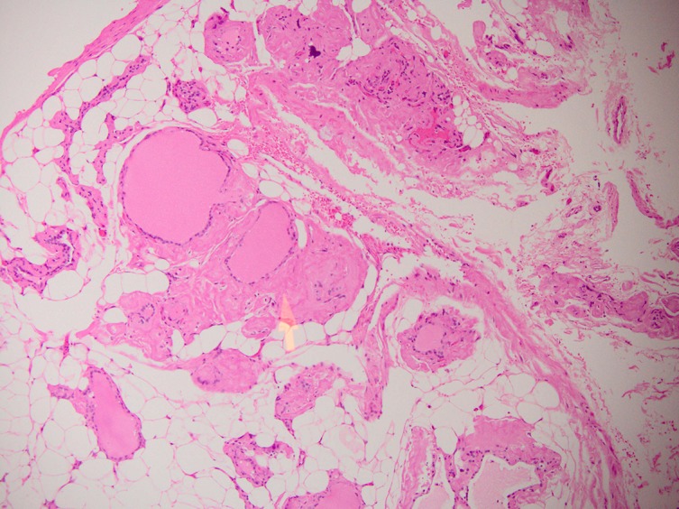
H+E stained section of the thyroid parenchyma not involved by cancer. Diffuse adipose infiltration of the parenchyma is seen. The yellow arrow shows amyloid protein deposition.

H+E stained section of the thyroid parenchyma not involved by cancer. Diffuse adipose infiltration of the parenchyma is seen. The yellow arrow shows amyloid protein deposition.