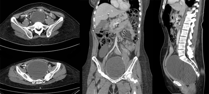
Figure 2.

CT imaging. On the left, axial slices showing displaced rectum (arrow) and uterus (asterisk). On the middle, coronal slice showing high extension up to iliac crests. On the right, sagittal slice showing relation with sacral bone and extension above the promontory. Ovarian cyst is not shown in this slice.
