
Figure 3.
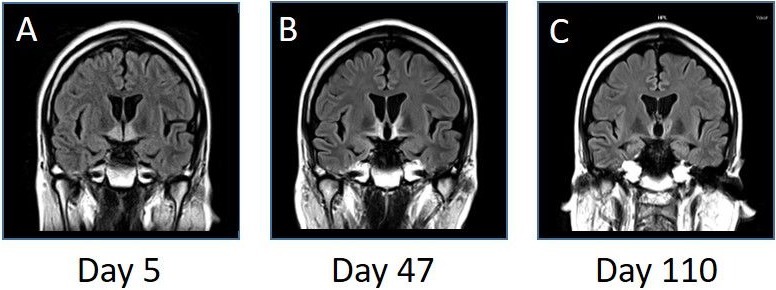
MRI (fluid-attenuated inversion recovery images, coronal, 3.0 T; TR 10 000 ms, TE 104 ms) on the fifth (A), 47th (B) and 110th (C) day after admission. The bilateral hypothalamic and anterior thalamic high-intensity lesions gradually improved on the following MRI. T, Tesla; TE, echo time; TR, repetition time.