Figure 2.

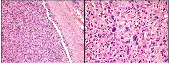
Well-defined intramuscular tumour composed of sheets and fascicles of highly pleomorphic spindle cells, cellular areas and myxoid background (H&E 100x and 400x).

Well-defined intramuscular tumour composed of sheets and fascicles of highly pleomorphic spindle cells, cellular areas and myxoid background (H&E 100x and 400x).