Fig. 4.
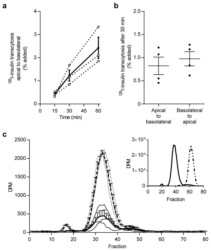
125I-TyrA14-insulin (125I-insulin) is transported across iBECs and remains intact. 125I-TyrA14-insulin transcytosis over 60 min (a). Dotted lines are individual experiments (n=3) and solid line is mean ± SEM. 125I-TyrA14-insulin transcytosis after 30 min is bidirectional and directions did not differ significantly (b). Data are presented as means ± SEM, paired t test. Basolateral media radioactivity (disintegrations per min, DPM) from transwell experiments sampled 30 min after apical 125I-TyrA14-insulin addition eluted from a Sephadex G-50 column in the same fractions in presence (solid line) or absence (dashed line) of iBECs (c). Thin dotted lines represent individual experiments without iBECs and thin solid lines represent individual experiments with iBECs (n=3). These peaks corresponded to intact insulin (insert, solid line) and did not appear in the same fraction as HepG2-degraded insulin (insert, dotted line)

