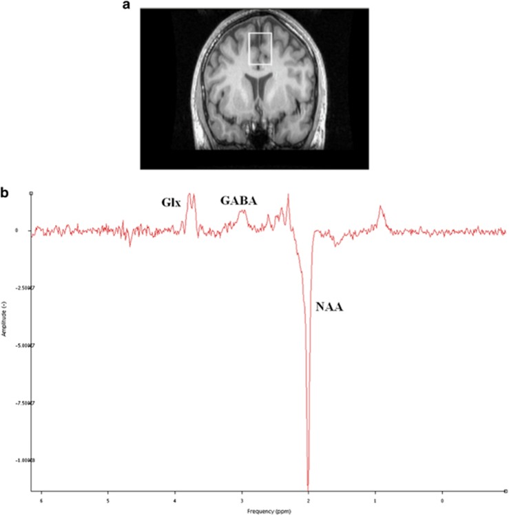
Figure 1.
[1H]MRS voxel positions and example spectra. (a) Cortical region of interest; medial prefrontal cortex (mPFC) (25 × 30 × 40 mm3) outlined in white, comprising primarily of anterior cingulate cortex. (b) Example spectroscopy output from the prefrontal voxel (Glx (glutamate+glutamine); GABA; N-acetylaspartate (NAA) identified using jMRUI spectroscopy software.

