Figure 5.
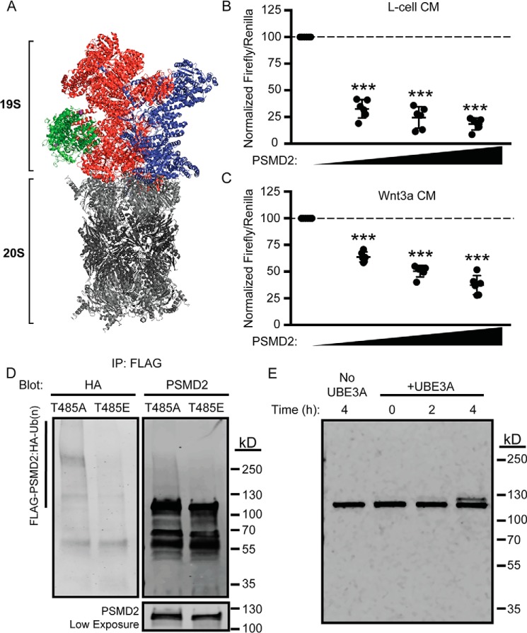
PSMD2 is a substrate of UBE3A. A, cryo-EM structure of the proteasome showing the location of PSMD2 (green) in the 19S regulatory particle (Protein Data Bank code 5GJQ). B and C, PSMD2 dose-dependently rescues UBE3A-stimulated Wnt signaling. HEK293T cells were co-transfected with plasmids encoding UBE3AT485A and increasing quantities of PSMD2 (0, 20, 40, and 60 ng). Cells were then grown in L-cell CM (B) or Wnt3a CM (C). Mean percent values for firefly:Renilla ratios are shown relative to cells transfected with UBE3A + empty vector (n = 6). Error bars indicate S.D. ***, p < 0.0005, one-sample t test (two-tailed). D, HEK293T cells transfected with the indicated UBE3A, Myc-DDK-PSMD2, and HA-ubiquitin constructs were treated with the proteasome inhibitor MG-132 (30 μm; 4 h). PSMD2 was immunoprecipitated (IP) using an anti-FLAG antibody, and the Western blot was probed with an anti-HA antibody (left panel) or PSMD2 antibody (right panels) to detect ubiquitinated PSMD2. E, an in vitro ubiquitin assay was performed using recombinant E1, E2 (UBE2D3), UBE3A, and PSMD2 expressed and purified from HEK293T cells. Reactions were stopped at the indicated time, and the formation of ubiquitinated PSMD2 was monitored using a PSMD2 antibody. WT UBE3A was omitted from the reaction as a negative control (first lane).

