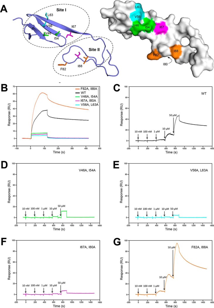
Figure 4.
Mutational analysis of the BMP-2-binding epitope of Col2a vWC. A, ribbon diagram of the Col2a vWC crystal structure showing surface-exposed hydrophobic residues selected for mutagenesis in site I and II. B–G, binding of Col2a vWC variants to immobilized BMP by SPR analysis. B, SPR sensorgrams of 40 μm wild-type (black) and mutants (colored as in A) of Col2a vWC binding to BMP-2. C–G, SPR sensorgrams of increasing concentrations of Col2a vWC variants (colored as in A and B) ranging from 10 nm to 50 μm sequentially injected at the time points indicated by arrows. RU, response units.

