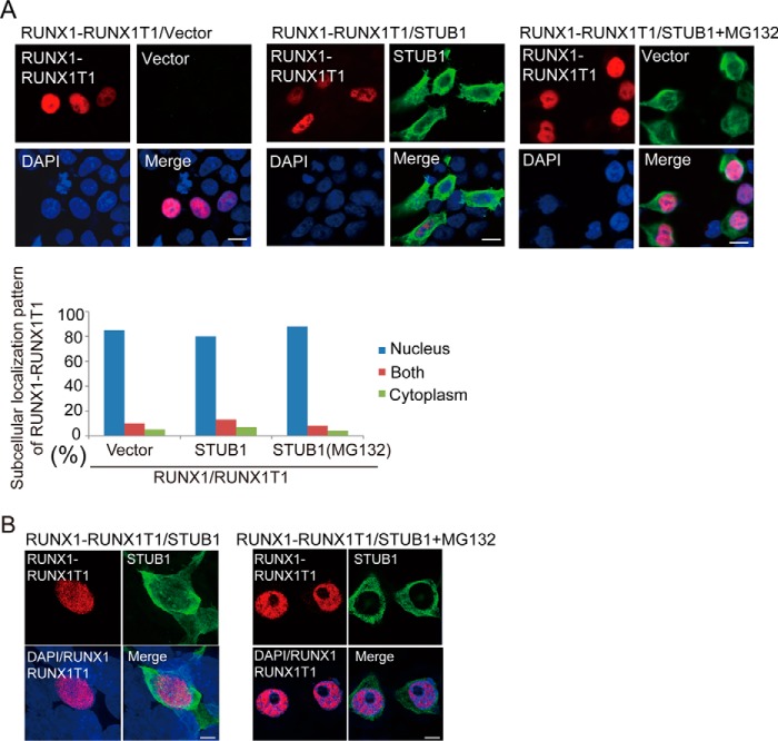
Figure 8.
STUB1 did not promote nuclear export of RUNX1–RUNX1T1. A and B, 293T cells were cotransfected with HA-tagged RUNX1–RUNX1T1 together with vector or FLAG-tagged STUB1 in the presence or absence of MG132 (20 mm), and were stained with anti-RUNX1 (rabbit) and anti-FLAG (mouse) antibodies followed by anti-rabbit Alexa 568 (red) and anti-mouse Alexa 488 (green) staining. Nuclei were visualized with DAPI (blue). Confocal laser scanning microscopy (Nikon A1, bar, 10 μm) (A) or super resolution microscopy (Nikon SIM, Bar, 5 μm) (B) was used to observe localization of RUNX1–RUNX1T1 and STUB1. STUB1 expression did not alter the subcellular localization of RUNX1–RUNX1T1.

