Figure 6.
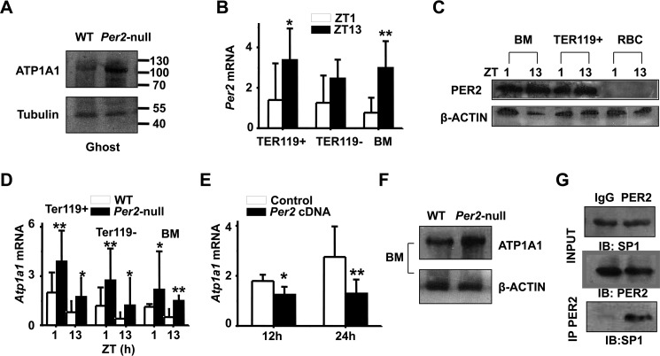
PER2 interacts with SP1 to repress Atp1a1 transcription. A, ATP1A1 protein levels in WT and Per2-null RBC ghosts. Representative blotting shows increased ATPA1 protein level in RBC ghosts of Per2-null mice. B, bone marrow erythroblasts were fractionated according to the developmental marker Ter119. Ter119+, Ter119−, and total bone marrow (BM) cells from wild-type mice were analyzed for Per2 mRNA expression at ZT1 and ZT13. C, Western blot analysis showing PER2 protein expression in BM and Ter119+ cells at ZT1 and ZT13 in WT mice. D, increased mRNA expression of Atp1a1 in Per2-null Ter119+, Ter119−, and BM. E, quantitative real-time RT-PCR analysis reveals a decline in Atp1a1 mRNA in TF-1 cells transfected with Per2 cDNA plasmid for 12 and 24 h. All values are mean ± S.D. (error bars); n = 6 in each group;*, p < 0.05; **, p < 0.01. F, Western blot profiles of ATP1A1 in BM and the RBC ghosts of WT and Per2-null mice. G, PER2 was immunoprecipitated from mouse BM. Total lysates (INPUT) and immunoprecipitation samples were analyzed by immunoblotting (IB) with SP1 antibody. Shown are representative pictures of three independent experiments.

