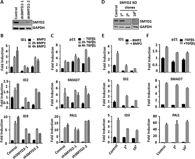
Figure 2.
Validating the role of SMYD2 in regulating BMP but not TGFβ target gene expression. A, Western blot analysis showing substantially reduced levels of SMYD2 in HaCaT cells stably infected with two different lentivirus-based shRNAs (shSMYD2-1 and -2). B, shRNA-infected HaCaT cells were serum-starved overnight and then stimulated with BMP2 for 0, 2, and 4 h as indicated. The cells were harvested, and qRT-PCR was performed to detect BMP downstream genes ID1, ID2, and ID3. C, shRNA-infected HaCaT cells were serum-starved overnight and then stimulated with TGFβ1 for 0, 2, and 4 h as indicated. The cells were harvested, and qRT-PCR was performed to detect TGFβ downstream genes p21, SMAD7, and PAI1. D, Western blot analysis showing the absence of SMYD2 proteins in SMYD2 knock-out HaCaT cell lines 2 and 16. The HaCaT knock-out cell lines were generated via CRISPR/CAS9 approach as described under “Experimental procedures.” E, SMYD2 KO 2 and 16 cell lines were serum-starved overnight and then stimulated without or with BMP2 for 4 h as indicated. The cells were harvested, and qRT-PCR was performed to detect BMP downstream genes ID1, ID2, and ID3. F, SMYD2 KO 2 and 16 cell lines were serum-starved overnight and then stimulated without or with TGFβ1 for 4 h as indicated. The cells were harvested, and qRT-PCR was performed to detect TGFβ downstream genes p21, SMAD7, and PAI1.

