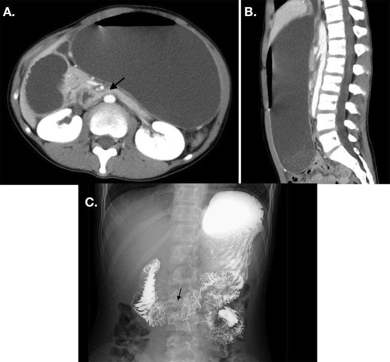
Figure 1.
(A) CT axial section shows a sandwiched horizontal portion of the duodenum between the superior mesenteric artery (SMA) and abdominal aorta (arrow), which was was proximal to the duodenal and gastric dilation. The distance between the aorta and SMA is 5.2 mm. (B) CT sagittal section shows a 17° angle between the aorta and the SMA. (C) An upper gastrointestinal series shows an abrupt cut-off (arrow) at the horizontal portion of the duodenum.

