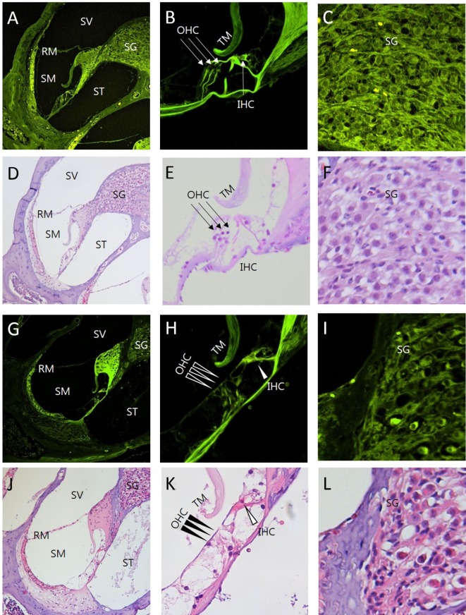
Figure 5.
Comparison between intravital multiphoton microscopy (MPM) and conventional light microscopy in the sections of the cochlea exposed to ototoxicity. We compared the intravital MPM images without any staining in the cryosections from normal mice (A–C) and mice exposed to ototoxicity (G–I) with hematoxylin and eosin (H&E) staining of normal mice (D–F) and mice exposed to ototoxicity (J–L). The images acquired after H&E staining and the intravital MPM images reveal similar morphological changes caused by ototoxicity in the cochlea, organ of Corti, and spinal ganglion. The organ of Corti (B,E,H,K) shows a clear cytoarchitecture. Note the appearance of the organ of Corti in a normal mouse (B,E) and the injuries to the IHC in the basal turn (H,K): the arrows indicate the area without supporting cells, pillar elements, or hair cells [(H,K) IHC, open arrowheads; OHC, arrowheads]. Note the appearance of the SG in a normal mouse (C,F). In contrast, the nerve fibers show diminished staining and decreased neuronal packing density in the SG (I,L) of a mouse exposed to ototoxicity. Intravital MPM can be successfully used to image the cochlea, and the cochlear structures show good correlation between the intravital MPM images without any staining and conventional light microscopic images with H&E staining. SV, scala vestibuli; SM, scala media; ST, scala tympani; RM, Reissner’s membrane; TM, tectorial membrane; BM, basilar membrane; SG, spiral ganglion; OHC, outer hair cells; IHC, inner hair cells.

